Neonatal Respiratory Distress

Learning outcomes
Introduction
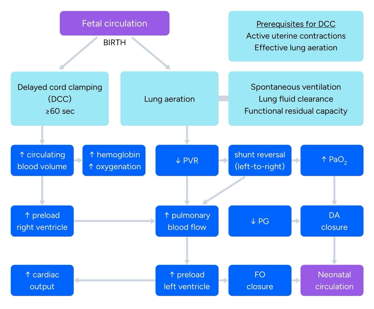
Successful postnatal transition depends on a rapid cascade of physiological milestones:
When any part of this process falters, neonatal respiratory distress develops—clinically defined by any change to frequency and/or quality of breathing pattern (i.e. abnormal breathing rates or increased work of breathing). A normal neonatal respiratory rate is 40-60 breaths/min, characterised by quiet and unlabored breathing.
However, during the first hours after birth (the immediate postnatal adaptation period), transient rates up to 60-70 breaths/min can occasionally be observed without distress, but rates consistently >60 breaths/min (tachypnea) or accompanied by signs of respiratory distress signal pathology.
Postnatal adaptation
Spontaneous respiratory effort and the establishment of FRC drives a rapid rise in arterial partial pressure of oxygen (PaO2) in the pulmonary vasculature (3 kPa → 10 kPa) within minutes, dramatically reducing pulmonary vascular resistance (PVR).
Together with the reversal of ductal shunting across the ductus arteriosus (right-to-left → left-to-right), this markedly enhances pulmonary perfusion, left ventricular preload, and cardiac output. These physiological transitions can be further augmented by delayed cord clamping (DCC), which increases circulating blood volume, improves myocardial contractility, and optimizes tissue oxygen delivery.
When postnatal adaptation is impaired, neonatal respiratory distress develops—defined as an alteration in the character and/or frequency of breathing. Neonatal respiratory distress remains one of the most common indications for neonatal intensive care unit (NICU) admission.
While often apparent immediately after delivery, symptoms may also develop later during hospitalization or following discharge home. For practical and didactic clarity, etiologies of neonatal respiratory compromise are broadly classified into pulmonary and extra-pulmonary causes.
Risk factors
Diagnosis
Physical examination (inspection, auscultation) and the measurement of peripheral oxygen saturation via pulse oximetry (SpO2) form the cornerstone of initial assessment. In the immediate postnatal period, the pulse oximeter probe is preferentially placed on the right upper extremity (wrist or palm) to measure preductal saturation.
Clinical findings
Typical signs of neonatal respiratory distress:
Newborns with respiratory distress relatively often present with circulatory or systemic signs:
Laboratory investigations
Imaging modalities
Differential diagnosis
Pulmonary causes of neonatal respiratory distress:
Extra-pulmonary causes of neonatal respiratory distress:
Therapy
General therapy
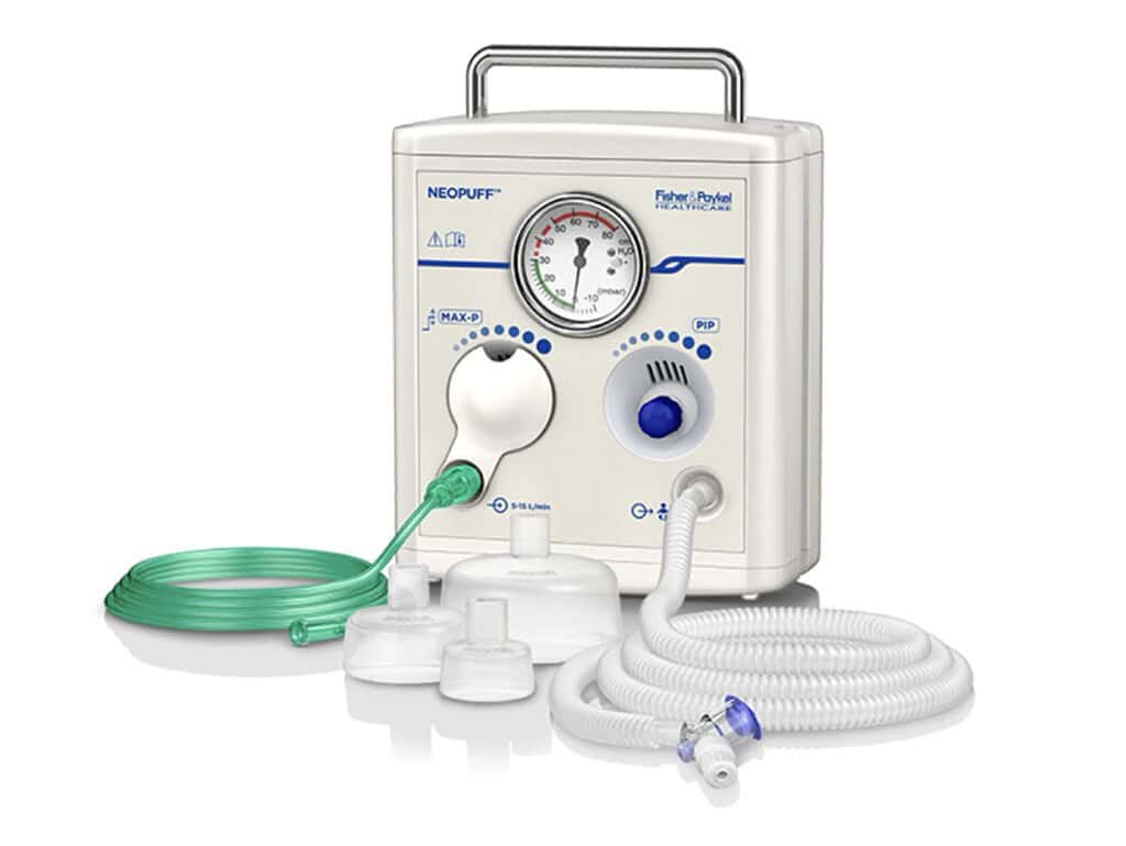
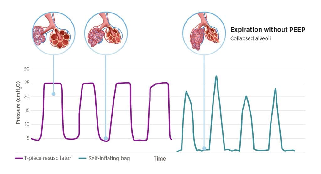
Currently, when a newborn experiences compromised postnatal adaptation with signs of respiratory distress, a T-piece resuscitator (e.g., Neopuff) is most commonly used, offering several advantages over conventional bag-valve-mask resuscitation. Compared to self-inflating and flow-inflating bags, T-piece resuscitators provide better-controlled, more consistent peak inspiratory pressure (PIP) alongside reliable positive end-expiratory pressure (PEEP), thereby helping protect the neonatal lungs.
Specific therapy
Z hlediska specifické terapie dechové tísně se používá:
🟨 surfaktant (Curosurf) – u nezralých novorozenců nebo u dětí se sekundární inaktivací surfaktantu (aspirace mekonia, plicní apoplexie, adnátní pneumonie)
🟨 kortikosteroidy (hydrokortizon, dexamethason, budesonid) – zlepšení funkce plic (systémové nebo inhalační podání)
🟨 bronchodilatační terapie (salbutamol, terbutalin)
🟨 hrudní drenáž (pneumothorax)
🟨 chirurgická intervence (vrozené srdeční vady, atrézie jícnu, vrozená brániční hernie)
🟨 parenterální nutrice (závažný stupeň dechové tísně, hypoglykémie, vrozené srdeční vady)
🟨 antibiotika (infekce)
🟨 terapie plicní hypertenze (oběhová podpora, analgosedace, inhalační oxid dusnatý)
🟨 dechová rehabilitace (dlouhodobá UPV)
- Oxygen (to reduce hypoxic injury, esp. brain; careful with overdosing = hyperoxia = retinal damage); pulse oximetry used to monitor hemoglobin oxygen saturation; oxygen (gas) needs to be warmed and humidified; FiO₂ 0.21 = 21% oxygen concentration = air; FiO₂ 1.0 = 100% oxygen concentration; vital signs monitoring – temperature, blood pressure, saturation, heart rate)
- Ventilation support (CPAP = non-invasive; mechanical ventilation = invasive)
- Surfactant (causal therapy; animal surfactant from pig lungs; synthetic surfactant; needs to be warmed up before bolus administration; immediate effect = increased lung compliance)
- Circulation support (severe RDS, sepsis, PPHN = inhaled nitric oxide)
- Parenteral nutrition (energy requirements, antioxidants)
- Caffein, Antibiotics, Analgesia/Sedation (during mechanical ventilation = blocks interference)
- Surgery if necessary (cleft palate, diaphragmatic hernia, congenital airway malformations, oesophageal atresia, congenital heart defects)
- Nursing care (secretions, handling, vitals signs monitoring, comfort and pain scoring, invasive lines check-up – endotracheal tube, cannulas, skin care, enteral feeding, eye care)
- Home oxygen therapy (BPD)
- Prevention of RSV infection (Synagis = Palivizumab = passive immunisation)
Pulmonary causes
Hyaline membrane disease (HMD)
- Typical for very low birth weight (VLBW) infants (birth weight < 1500 grams)
- Quickly develops during the first hours of life = requires complex treatment (see Therapy)
- Uncomplicated HMD lasts usually 3-5 days
- a hyaline membrane composed of proteins (intra-alveolar leak) and dead cells inside the alveoli (pathologic histology of lungs of premature newborns)
- anatomical and functional prematurity of lungs (i.e. typical for preterm newborns)
→ anatomical = preterm lungs in saccular developmental stage (abundant interstitial tissue and insufficient inner spacing)
→ functional = inability to maintain residual volume due to surfactant deficiency => focal atelectasis surrounded with areas of hyperinflation => reticular granular pattern on chest X-ray (+ intra-alveolar protein leak from plasma to alveoli causing edema and surfactant dysfunction)
Surfactant
- surface active agent
- prevents alveoli from collapse at the end of expiration
- produced and recycled by pneumocytes II during the second half of pregnancy
- composed of 90% phospholipids (phosphatidylcholine = lecithin + phosphatidylglycerol) and 10% proteins (SP-A/B/C/D)
Transitory Tachypnea
- “wet lung” syndrome = caused by diminished pulmonary fluid resorption
- preterm and term newborns (cesarean delivery, maternal diabetes, perinatal asphyxia)
- may require complex therapy, however, usually resolves within 12-24 hours (compared to HMD)
Meconium aspiration syndrome (MAS)
- fetal hypoxia = gasping = amniotic fluid aspiration => meconium in the fluid = meconium aspiration syndrome
- requires support right after delivery (chest X-ray with focal atelectasis or decreased transparency)
- meconium in lungs = inflammation, surfactant dysfunction, PPHN = respiratory failure = mechanical ventilation, surfactant, inhaled nitric oxide
Pneumonia
- intrauterine / perinatal aspiration of infectious amniotic fluid (chorioamnionitis)
- Streptococcus agalactiae (Group B Streptococcus = GBS), Gram negative bacteria, Ureaplasma spp.
- lung inflammation = surfactant dysfunction, PPHN + sepsis (BSI = blood-stream infection) = respiratory-circulatory failure = mechanical ventilation, surfactant, inhaled nitric oxide, antibiotics
Air leak syndromes
- air leak to interstitial tissue (PIE = pulmonary interstitial emphysema) or pleural cavity (PNO = pneumothorax)
- asymmetrical auscultation + typical chest X-ray findings
- Spontaneous = vigorous breathing following amniotic fluid aspiration; can resolve spontaneously
- Iatrogenic = artificial ventilation / mechanical ventilation; can cause tension PNO
- tension pneumothorax is an emergency presenting with sudden bradycardia and/or desaturations and requires chest drainage (unilateral x bilateral with FR 8/10 tube inserted in the 3rd intercostal space) !
Bronchopulmonary dysplasia
- chronic lung disease (BPD) caused by various factors (infection, degree of prematurity, nutrition, aspiration, oxygen, mechanical ventilation, fetal growth restriction) on immature and developing lung tissue (preterm newborns!) = anatomical and functional changes
- alveolar space restriction + interstitial tissue increase + disruption of alveolarization and vascular bed formation
- mild BPD = oxygen dependency at day of life (DOL) 28
- moderate BPD = oxygen dependency at 36 weeks of gestation (36+0)
- severe BPD = mechanical ventilation at 36 weeks of gestation (36+0)
- Chest X-ray : lung fibrosis, heterogenous involvement, focal emphysema and atelectasis, diffusely decreased transparency
- Treatment (see Therapy)
- Eventual resolution – oxygen and functional rejuvenation within 1 year (usually)
- Pulmonary hypertension (cor pulmonale) – worse prognosis
References
① Sweet DG, Carnielli V, Greisen G, et al. European Consensus Guidelines on the Management of Respiratory Distress Syndrome – 2019 Update. Neonatology. 2019;115(4):432-450. doi:10.1159/000499361
② Wang C, Guo L, Chi C, et al. Mechanical ventilation modes for respiratory distress syndrome in infants: a systematic review and network meta-analysis. Crit Care. 2015;19(1):108. Published 2015 Mar 20. doi:10.1186/s13054-015-0843-7
③ Dargaville PA, Gerber A, Johansson S, et al. Incidence and Outcome of CPAP Failure in Preterm Infants. Pediatrics. 2016;138(1):e20153985. doi:10.1542/peds.2015-3985